Phage Biology: The Life of Phage
Bacteriophages, viruses that infect and kill bacteria, are the most numerous entities on the planet. Yet, despite how abundant and prevalent phages are, these tiny bacteria-parasitizing virions weren’t discovered until the early 1900s. Since then, we have learned a lot about the fundamentals of phages, like what they are and how they work. To this day, scientists continue to explore the immense diversity of phages.
Mysterious antibacterial activity
At the turn of the 20th century, scientists initially noticed bacteriophages for their ability to kill bacteria, but it wasn’t immediately clear what they had found. They recorded cases where certain water or fecal filtrates could inhibit the growth of bacteria, which is now referred to as the “bacteriophage phenomenon.” Early hypotheses debated whether it was a protein, a chemical, a virus, or some other substance responsible for the observed bacterial death. Without knowing exactly what he was working with, Félix d’Hérelle, a French-Canadian microbiologist, called the mysterious bacteria-eating microbes “bacteriophages,” a name that stuck.
Over the following decades, researchers continued to characterize phage behavior and applications. Finally, in 1940, German scientists had a powerful enough microscope to see what was eliminating bacteria—these tiny, peculiar bacteriophage particles. With time, the phage photos circulated among scientific communities across the globe.

Taking a closer look at phages
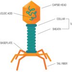
Like other viruses, phages are not cellular. Instead, they have genetic material inside a protein shell called a capsid head.
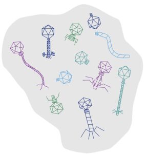
Phages have immense biodiversity. They are typically categorized based on the type of genetic material they have and their morphology (how they appear under a microscope).

Depending on the phage type, the genetic information can be deoxyribonucleic acid (DNA) or ribonucleic acid (RNA), either single or double-stranded. To date, a majority of characterized phages have DNA genomes.
Over 96% of known phages are Caudovirales, or “tailed viruses.”(1) Giving phages the look of a lunar lander, the tail is an appendage stemming off the capsid head capped with little leg-like projections called tail fibers.(2) The most studied families of tailed phages include Myo-, Sipho-, and Podoviridae types with varying tail morphology. Myoviridae have a long contractile tail, while Siphoviridae and Podoviridae have noncontractile tails that are long and short, respectively. (What type of phage is shown under the microscope in the image above? Keep reading to find out!)

The capsid head of tailed phages usually appears spherical at a glance, but many are actually icosahedral shaped with 20 triangular faces. Notably, capsid structures are often symmetrical, with repeating protein subunits on each side. These highly ordered protein shells house and protect the phage’s genetic information. In fact, DNA packed tightly inside can actually expand the capsid a bit.
Other phages like Corticoviridae and Microviridae have semi-spherical capsids without any tail. Filamentous phages (Inoviridae) are also tailless but with a long wormy-shaped capsid. Interestingly, fifteen types of non-tailed phages are known, yet they make up only 4% of inspected phages.1 It is still unclear to scientists why this might be, but they wonder if tailed phages are most easily recognized under the microscope or if the tail has some major benefits for the phages (more below on what the tail is for).
Over the past 80 years, scientists have primarily relied on morphological traits to classify phages. However, in recent years, advancements in DNA sequencing technology have allowed for an even closer inspection of phages and more detailed characterization. Genetic analysis has revealed a rather complicated web of evolutionary relationships among phages. Interestingly, even phages that appear similar at a glance can have wildly different genetic markers, suggesting they are not as closely related as initially thought. Scientists continue to refine the classification system of phages as more are discovered and investigated.
Enter phage: how bacteria get infected
Like other viruses, phages are non-living entities that cannot reproduce independently; they depend on bacterial hosts to replicate and survive. To expand their populations, phages must infect bacteria.(3)
Phages are very particular about what bacterium they will parasitize. Each phage type has a narrow host preference, usually only select species or even strains of bacteria. For example, one of the most well-characterized phages, called T4, exclusively infects Escherichia coli.
To achieve this remarkable specificity, bacteriophages use an elaborate “lock and key” recognition system to identify bacterial cells of interest. That is, a phage recognizes and binds the target microbe through specific molecules that coat the bacterium.
Various proteins and other molecules on the outside of the bacterium make it recognizable to its phage predator including polysaccharides, outer membrane proteins, and components of flagella and pili structures, to name just a few.
To detect and discern their prey, phages use so-called receptor binding proteins (RBPs) at the phage’s tail-end. Some tails have intricate structures at the tip, including baseplates, spikes, and fibers to guide specific binding to the bacterial host.
In fact, host recognition is a complex process where many tail proteins interact with multiple different surface molecules on the bacteria. For example, Escherichia coli is targeted through lipopolysaccharide (LPS) and OmpC porin proteins by the T4 phage.(4) On the other hand, a siphophage called iEPS5 attaches to Salmonella by recognizing FliC and FljB proteins of the bacterium’s flagellum propeller.(5) These redundant mechanisms ensure the correct bacterium is selected and infected to replicate the phage successfully.
Once a phage finds and attaches to a suitable bacterial cell, the infection process continues. Enzymes on the tip of the phage’s tail eat away at the exterior of the bacteria. These same types of enzymes allow phages to degrade and infiltrate biofilms, the tough tight-knit bacterial colonies that can contribute to antibiotic resistance.
After digging through the outside layer, phages inject their viral genetic material into a bacterial cell. In addition to facilitating recognition, the tail is also a conduit connecting the virus to the target microbe. The phage’s genetic material passes through the tubular tail and enters the bacterium.
To kill or not to kill: lytic or lysogenic phages
What happens once a phage has injected its DNA (or RNA) into a bacterium? Very rapidly, the bacterial cell turns into a phage manufacturing plant. The cellular machinery is hijacked to produce new phage components that quickly assemble to form new phages. When they get the signal, the fresh phages burst the bacterium. Phage multiplication happens in minutes to hours, and over a thousand viral particles spew from the dead bacterium. Other bacteria in the immediate area are now at risk of being infected next.

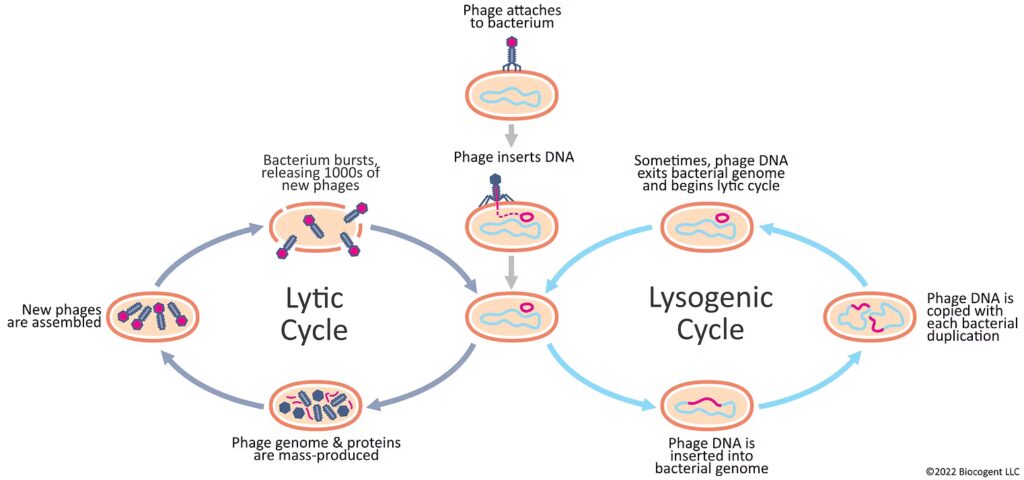
Virulent phages reproduce through this deadly process that is known as the “lytic cycle.” Along with generating many new phage particles, the lytic cycle also breaks open (or lyses) the cell, resulting in the death of that bacterium. Notably, therapeutic applications of phages, commonly referred to as “phage therapy,” depend on the lytic cycle to rapidly kill infected bacteria.
For this reason, so-called temperate phages that follow the “lysogenic cycle” are not typically useful for antibacterial applications. Instead of killing microbes, temperate phages will infect bacteria and enter a dormant state. The genetic material quietly integrates into the bacterial genome instead of starting phage replication. In most cases, the bacterial cell is not affected by the infection of a temperate phage and will continue to grow as usual. The dormant phage genes may eventually reactivate in certain circumstances to produce phage particles.
Key points
- Phages have immense biodiversity. Scientists continue to discover and investigate new phages.
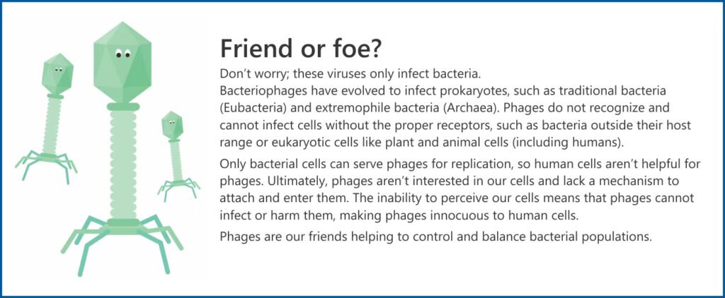
- Phages are highly specific with what cells they will parasitize. They don’t recognize or infect human cells and are very selective in the species or strain of bacteria they will choose.
- Virulent phages reproduce through the lytic cycle that rapidly kills the bacteria. In the lysogenic cycle, temperate phages go dormant and don’t usually kill the bacteria.
Still wondering?
The phages shown under the microscope are Siphoviridae! These are the long, non-contractile-tailed bacteriophages found in DermaPhage® CA .
References
- 1 Ackermann, H. W. 5500 Phages examined in the electron microscope. Archives of Virology 152, 227-243, doi:10.1007/s00705-006-0849-1 (2007).
- 2 Dion, M. B., Oechslin, F. & Moineau, S. Phage diversity, genomics and phylogeny. Nature Reviews Microbiology 18, 125-138, doi:10.1038/s41579-019-0311-5 (2020).
- 3 Stone, E., Campbell, K., Grant, I. & McAuliffe, O. Understanding and Exploiting Phage-Host Interactions. Viruses 11, doi:10.3390/v11060567 (2019).
- 4 Washizaki, A., Yonesaki, T. & Otsuka, Y. Characterization of the interactions between Escherichia coli receptors, LPS and OmpC, and bacteriophage T4 long tail fibers. Microbiologyopen 5, 1003-1015, doi:10.1002/mbo3.384 (2016).
- 5 Choi, Y., Shin, H., Lee, J. H. & Ryu, S. Identification and characterization of a novel flagellum-dependent Salmonella-infecting bacteriophage, iEPS5. Appl Environ Microbiol 79, 4829-4837, doi:10.1128/aem.00706-13 (2013).